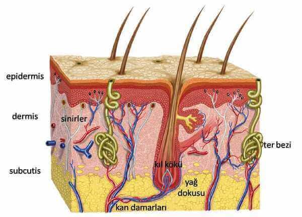
Our skin (skin) is the largest organ after the skeletal system. The surface area is approximately 1.9 m2 per person covers the entire body. Mouth, eye genital area as mucous structure continues.
The weight of our skin is about 15% of the body weight of an adult.
Although it varies in different parts of our body 1 cm2 area of our skin; on average 10 hair follicles, 15 sebaceous glands, 100 sweat glands, half meter blood vessel, 2 meters nerve, 3000 sensory nerve endings, 200 pain-sensing nerve endings, 25 pressure-sensing nerve members, 2 cold sensing nerve s 12 The pieces are hot-sensing nerve member.
How is the Skin / Skin Appearance?
When we look from the outside, we see thin thick lines hairs pores on the outer surface of our skin that form structurally crossed square, rectangular, triangular trapezoidal shapes.
The geometric shapes formed by the thin thick lines we see on our skin are called “sulcus cutis hieroglyphic patterns.. The thick lines, called primary lines, are broad deep. The thin lines called secondary lines are shallow narrow. These lines shapes vary according to body parts.
Apart from these structural lines on our skin, there are also wrinkles caused by deformation of the skin due to aging, mimic sleep patterns. Subsequent wrinkles on our skin are in two forms: superficial deep.
Superficial wrinkles are non-deep wrinkles, which are caused by the loss of water in the epidermis layer the deterioration of the epidermis.
Deep wrinkles are caused by degradation of the dermis due to loss of elasticity due to decrease in collagen elastin fibers.
The hairs on our skin are the product of a structure called hair root. The distribution of hairs is genetically determined the hair roots are found in all parts of our skin except the soles of the feet the palms. While these hair roots produce small thin hairs that are too small to be seen with our eyes in some regions, they work actively in some regions.
The pores in our skin appear widely on the skin, showing the mouths of the channels the oil sweat glands open to the skin surface.
What is the Duty of Our Skin / Skin?
Our skin; Provides the relationship between our body the external environment, protects against internal external factors.
Duty against internal factors; detoxification regulation protection of body temperature.
The task of detoxification; toxins, water salt from the body by sweat.
It regulates maintains body temperature with the special structure of skin fat tissue skin vessels in hair sebaceous glands sweat glands.
The duty to protect against external factors;
-
Protection against biological agents,
The acid mantle system in our skin maintains the pH of the skin against bacteria between 4.2-5.6, the epidermis is constantly poured out of the skin, compact structure of the epidermis, realizes with rich defense system cells.
-
Protection against physical factors;Mechanical factors; the compact structure of the epidermis, collagen elastin bundles in the dermis, subcutaneous adipose tissue. Protection against light, heat electricity
-
Protection against chemicals; Keratin lipid mantle provide this. The lipid mantle protects the skin against fungi.
-
Absorption ejection properties;
The skin is a metabolically active member. It breaks down hormone, sterodi inflammation intermediates from the body. Xenobiotic substances break down drugs, pesticides, environmental industrial chemicals. It removes chemicals that are soluble in oil water.
The skin also provides the absorption of drugs. The absorption of drugs chemicals is mostly on the ovary skin, forehead around the ears.
-
Discharge of static electricity from the body
-
Vitamin production task; Vitamin D is made.
-
Support task; covers supports the underlying tissues with its flexible feature
-
Sensory function; It senses sensations such as heat, pressure pain.
-
Pigmentation production task; It protects the skin lower tissues against UV by melanin synthesis. Its role in the immunological system; It plays an active role in the skin defense system.
-
Warehouse duty; Carbohydrates, fat, water blood are stored in the skin.
Aesthetic look; provides the aesthetic appearance of a body as high as an organism.
What is the Anatomical Structure of our Skin / Skin?
Our skin consists of 3 layers: epidermis, dermis subcutaneous tissue-pannicle. The thickness of these 3 layers of skin skin varies according to the anatomical regions of our body. For example, the thinest area of the epidermis is our eyelids with 0.1 mm. The thickest places are hand foot soles with 1.5 mm. The dermis is the thickest skin of the back.
Epidermis
It is the outermost layer of the skin consists mostly of cells that we call keratinocytes. Does not contain vascular structures. Its thickness varies between 05–100 microns (600 microns for hand collar), although it varies according to the body area. In addition, the water content of the epidermis is a factor that changes its thickness. Apart from keratinocytes, Melanocytes, Langerhans Merkel cells are also present in the epidermis. Keratinocytes constitute 5% of the skin make protein called keratin in the protein structure in the cell.
The keratinocytes are divided into the lower layers, ie the dermis the adjacent layer, discarded into the upper layers. In the lower layers, live keratinocytes die in the uppermost layers are excreted from the skin. This process is called keratinization cycle-turnover. In a normal person, this process is 28 days.
The epidermis consists of 4 sub-layers;
Stratum basale
Stratum spinosum
Stratum granulosum
Stratum corneum
The Dermis
Under the epidermal layer is the second layer of the skin. Dermis varies according to the body area, but it is 2-4 mm thick. The intercellular supportive tissue fibroblast cells in the dermis, including nerve, vascular, lymphatic structures, sweat sebaceous glands, nail hair follicles. Compared to the epidermis, there are far fewer cells more fibers.
The main cells in the dermis are fibroblasts make the extracellular support matrix tissue of the dermis. This matrix structure consists of collagen, elastin reticular fibers. The dermis also hosts other cells. Macrophages, such as mast cells, are part of the body's defense system.
It is found in receptors with sensory nerves in the dermis. Merkel Meissner bodies (sense of touch, mostly located in the hands soles of the foot), Pacinian bodies (pressure sense, body weight to the stone anatomical areas genital area are more intense) Ruffini bodies (mechanical sense).
Collagen in the dermis provides skin tension. Elastin gives the skin its elasticity. These two are the fibrillary support tissue of the dermis. Apart from fibrils, proteoglycans, glycoperoteins, glycosaminoglycans, water hyaluronic acid are other support structures. Glucosaminoglycans are the prominent ones. These are forms of proteins bound to proteoglycans. Proteins are chondrotin sulfate, dermatan sulfate, keratin sulfate, heparan sulfate heparin. The most important proteoglycan is versican (providing skin tension) perlecan (located in the basal layer between epidermis dermis). Glycoproteins are laminis, matriline, fibronectin, tenascin. These regulate the attachment of cells, the displacement of cells, the relationship between cells.
Subcutaneous Tissue (Hypodermis)
It is the lowest layer of the association. It consists of the cells we call lipocytes. Lipocytes make small chambers called panniculus. Endocrine tasks turn androgens into estrogens. They make leptin provide a sense of satiety.

